Microsurgical Vasectomy Reversal
Robert U. Finnerty M.D. FACS
 In the past, the results were very poor for vasectomy reversal, because of the small size of the vas deferens and the tiny opening (lumen) inside it. Older reversal techniques, generally using low-power optical magnification or jeweler’s loops, simply removed the scarring from the vasectomy site and placed a few large sutures to approximate the ends. Because the upper and lower ends of the vas have such different diameters after vasectomy, the inside channel was poorly aligned. This allowed sperm to leak out, resulting in inflammation and scarring.
Success as a result was relatively rare, and even when sperm appeared after surgery, sperm counts were often very low and fertility was poor. Attempts to improve the success rates using techniques such as stents failed to improve the results. And when a secondary obstruction from an epididymal leak occurred, these non-microsurgical techniques offered no hope for success.
The answer to these problems arrived in the mid-1970’s with the development of very high magnification surgical techniques, called microsurgery. Initially used to repair tiny blood vessels and nerves, it quickly found application in infertility surgery. Using a specialized piece of equipment called an operating microscope, surgeons with microsurgical training use sutures (stitches) which are barely visible to the naked eye, working with magnification between 20-50x (compared to 2-4x with optical loops).
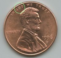 These microsurgical techniques allow extremely precise placement of sutures, resulting in a far more accurate alignment of the vas lumen and markedly improved success rates in vasectomy reversal surgery. Using microsurgery, eight or more sutures can be placed around the inside channel of the vas, which is the size of the “o” in “God” on a penny.